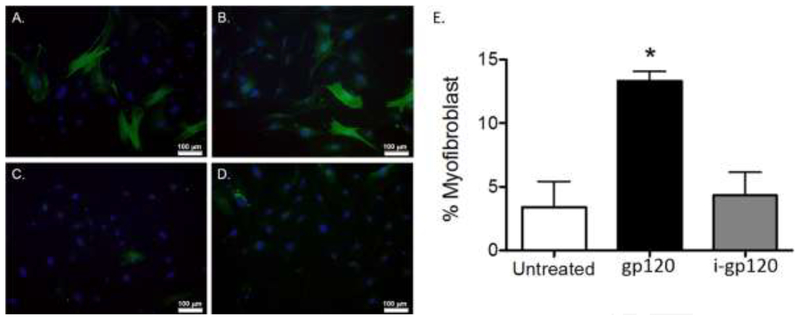
Figure 2. gp120 induces the myofibroblast phenotype in mouse primary lung fibroblasts.
Mouse primary lung fibroblasts (PLFs) were isolated from wild-type mice, and after passages 3–7, PLFs were treated with (A) gp120 (10ng/mL), (B) TGFβ1 (5ng/mL), (C) no treatment, and (D) heat-inactivated gp120 (10ng/mL). At 96 hours, cells were fixed and stained for α-SMA and DAPI. Cells were analyzed for morphology by immunofluorescence microscopy. (E) Graph depicts quantification of myofibroblasts by cell count. Total PLFs counted for untreated group = 222, gp120-treated group = 371, and heat-inactivated gp120-treated group = 328. Green = α-SMA, blue = DAPI nuclear stain. All images were captured at 20× magnification. *P<0.05 increased compared with untreated and heat-inactivated gp120-treated groups.

