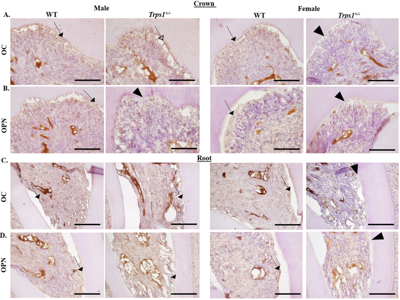
Figure 3. Immunohistochemical analysis of osteocalcin (OC) and osteopontin (OPN) in WT and Trps1+/− first mandibular molars of 4 wk old male and female mice.
(A, B) Representative images of sagittal sections of the crown portion of the molars. (C, D) Representative images of sagittal sections of the root portion of the molars. OC and OPN are detected in both crown and root odontoblasts and dentin of WT female and male mice (arrows). In Trps1+/− females, decreased expression of OC and OPN proteins was detected in both crown and root odontoblasts (arrowheads) in comparison with WT females. In Trps1+/− male mice, stronger OC signal was detected in crown odontoblasts and dentin (open arrowhead), while the expression of OPN was decreased (arrowhead) in comparison with WT males. Scale bars=100μm.

