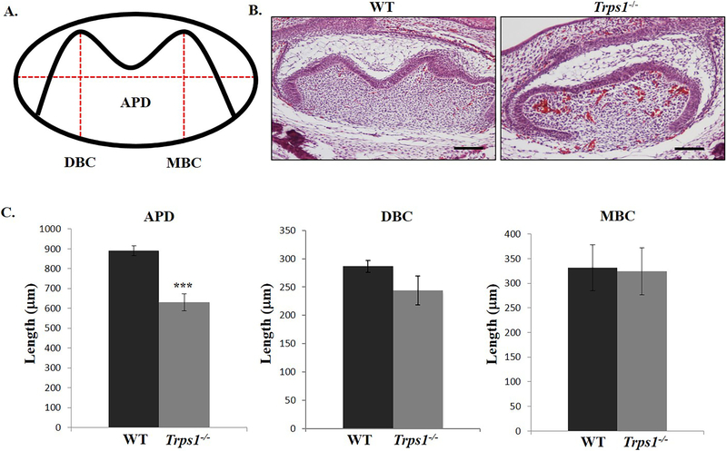
Figure 5. Comparison of the tooth organ dimensions (first mandibular molar) of E18.5 WT and Trps1−/− mice.
(A) Schematic of a tooth organ showing measurements of anterior-posterior diameter (APD), distal buccal cusp height (DBC) and mesial cusp height (MBC). (B) Representative images of H&E-stained sagittal sections at the midline of E18.5 tooth organs. Scale bar =250μm. (C) Graphs comparing APD, DBC height and MBC height of tooth organs of WT and Trps1−/− mice. Data are presented as mean values ± SD from equivalent sections (N=3 mice/genotype). ***p ≤0.0005.

