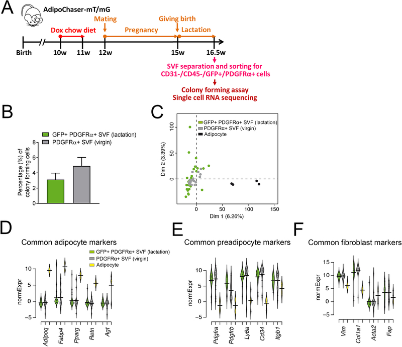
Figure 4. Characterization of de-differentiated adipocyte in the lactating mammary gland by single-cell RNA-seq.
(A) Experimental design: 10-week old AdipoChaser-mT/mG female mice were put on a doxycycline chow diet for 7 days to ensure that GFP expression is turned on in all mature mammary adipocytes. After doxycycline treatment, mice were switched back to regular chow diet for a week to ensure the washout of doxycycline. For chasing mature mammary adipocyte in lactating female, mice were bred with male mice to undergo pregnancy and lactation. When the mice had been lactating for 10 days, the mammary glands were used for SVF separation. The separated SVF was then flow-sorted for CD31−/CD45−/PDGFRα+/GFP+ cells for single-cell RNA sequencing analysis. (B) Quantification of colony-forming unit of FACS purified CD31−/CD45−/PDGFRα+/GFP+ from the mammary gland SVFs of lactating female, comparing to the classic preadipocyte population (CD31−/CD45−/PDGFRα+ cells) from the mammary gland SVFs of virgin female. (D-G) Gene expressions of these de-differentiated mammary adipocytes (CD31−/CD45−/PDGFRα+/GFP+ cells) from the mammary gland SVFs of lactating female were compared to the classic preadipocyte population (CD31−/CD45−/PDGFRα+ cells) from the mammary gland SVFs of virgin female and an adipocyte cell line through single-cell RNA-seq. (C) Overview of single-cell grouping by principal component analysis. (D-F) Beanplots showing the distribution of normalized expression values (normExpression) across cells that belong to one of these three categories of common adipocyte markers (D), common preadipocyte markers (E) and common fibroblast markers (F). n = 26 cells for CD31−/CD45−/PDGFRα+/GFP+ SVF cells from lactating female, n = 20 cells for CD31−/CD45−/PDGFRα+ SVF cells from virgin female, n = 4 cells for brown adipocyte cell line.

