Fig. 4.
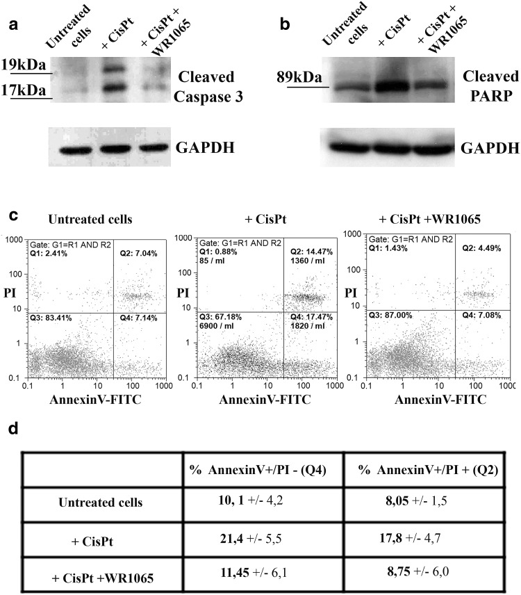
Analysis of apoptosis in NT2-N cells. Western blots (a, b) and flow cytometry analysis of apoptosis (c, d). For Western blots untreated cells and cells exposed to cisplatin alone (+ CisPt) or in combination with WR1065 (+ CisPt + WR1065) were harvested and their total protein content isolated. a Analysis of caspase 3 cleavage; activated Caspase 3 fragments are detected as 19 kDa and 17 kDa molecular weight bands. b Analysis of cleaved PARP; carboxy-terminal catalytic domain of PARP released upon cleavage generated 89 kDa band. GAPDH was used as a control of equal protein loading. c NT2-N cells were exposed to cisplatin alone or in combination with WR1065 and analyzed by flow cytometry after 24 h. Whole cells selected by gating were stained by PI and Annexin V and separated into quadrants. Q1: PI+/Annexin-cells are necrotic; Q2: PI+/Annexin + cells are late apoptotic cells; Q3: PI-/Annexin-cells are alive; Q4: PI-/Annexin V + cells are in early apoptosis. d Two independent flow cytometry experiments were done with similar outcomes—average values and standard deviation are presented in the table for early and late apoptotic cells

