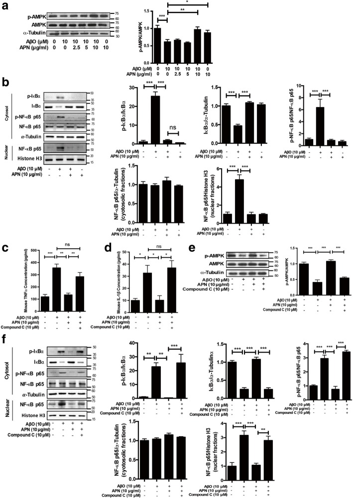
Fig. 3.
APN suppressed AβO-induced proinflammatory cytokine release in BV2 cells via AMPK-NF-κB signaling pathway. a Representative western blot analysis of AMPK phosphorylation. α-Tubulin immunoreactivity was used as a loading control (n = 6). b Representative western blot analysis of NF-κB activation in BV2 cells. The cytosolic fractions were prepared and analyzed with phosphorylated IκBα, total IκBα, phosphorylated NF-kB p65S536, and total NF-kB p65. The nuclear fractions were prepared and analyzed with total NF-kB p65. α-Tubulin immunoreactivity was used as a loading control in the cytosolic fraction, and histone H3 was used as the loading control in the nuclear fraction (n = 5). c, d BV2 cells were pretreated with AMPK inhibitor Compound C (10 μM) for 2 h, and then were treated with APN for 2 h, followed by exposure of AβO for 24 h. ELISA assays of TNFα and IL-1β were conducted (n = 3). e Representative western blot analysis of AMPK phosphorylation (n = 6). f Representative western blot analysis of NF-κB activation in BV2 cells (n = 5). Data were presented as the mean ± SEM. One-way ANOVA with Tukey’s multiple comparison test revealed a difference between groups. *p < 0.05, **p < 0.01, ***p < 0.001; ns, statistically not significant

