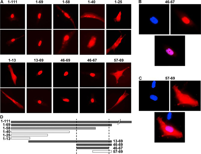
Figure 3.
Subcellular localization of the fusion constructs between the indicated segment peptides of FLIL33 and mScI. A. Fluorescence microscopy, red channel, of two representative cells for each FLIL33 segment is shown; repeated in primary NHLFs from three separate donors, with at least 50 overexpressing cells analyzed for each donor, with consistent results. B, C. Fluorescent images of cells with DAPI-stained nuclei (blue) expressing the indicated IL-33 segment peptide – mScI fusion proteins (red), and the overlays of the two channels. D. The horizontal bars represent the corresponding FLIL33 N-terminal peptide sequences to scale; closed bars represent nuclear and open bars extranuclear or combined localization.

