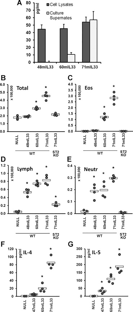
Figure 4.
Properties of N-terminal deletion mutants of mFLIL33. A. ELISA of cell lysates and cell culture supernates of primary mouse cells 72 h after electroporation with plasmids encoding the indicated mFLIL33 mutants, mean pg/ml ± SD. B – E. Total and differential (eosinophils, lymphocytes, neutrophils) cell counts of BAL samples from wild type and ST2 knockout mice infected intratracheally with AdV constructs encoding the indicated variants of mIL-33; day 10 after infection. F, G. ELISAs of lung homogenates for IL-4 (F) and IL-5 (G). In panels B – G, dots represent individual mice and horizontal bars show mean values for each group. Statistically significant differences (p < 0.05) from AdV-NULL-infected mice are indicated with asterisks.

