Figure 3:
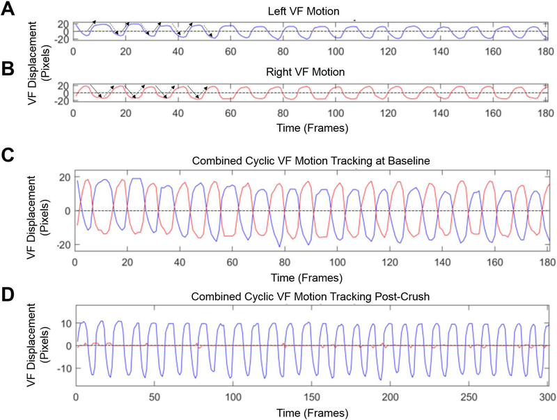
A & B) Displacement of the left (blue) and right (red) VFs individually over time. The x-axis is the video frame number and the y-axis is VF displacement measured in pixels. The graphs display the cyclic movement as the VFs oscillate back and forth across their respective motion midlines (dashed-lines) during inspiration and expiration in a normal mouse at baseline under a surgical level of anesthesia. Solid arrows indicate the VF is adducting (closing), whereas dashed arrows indicate the VF is in a state of abduction (opening). C) The left and right VF displacement graphs from baseline are overlaid with respect to their motion midlines. Right VF range and frequency of motion are similar to the left. D) Combined right and left VF movement immediately after a right RLN crush injury. There is no right VF movement, compared to normal left VF movement.

