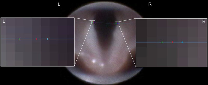
Figure 5:
Representative image of VFTrack validation process. Vocal fold (VF) boundaries were tracked with our automated software (red points). Manual points were placed on each frame by two independent reviewers (green and blue points). The expanded view of the left and right VF boundaries shows individual pixels and the pixel location of each point along the given blue horizontal line. In this image, both reviewers are no more than two pixels away from the automatically tracked point, demonstrating high reliability of our automated tracking software. For perspective, the total endoscopy field of view contains approximately 60,000 pixels.

