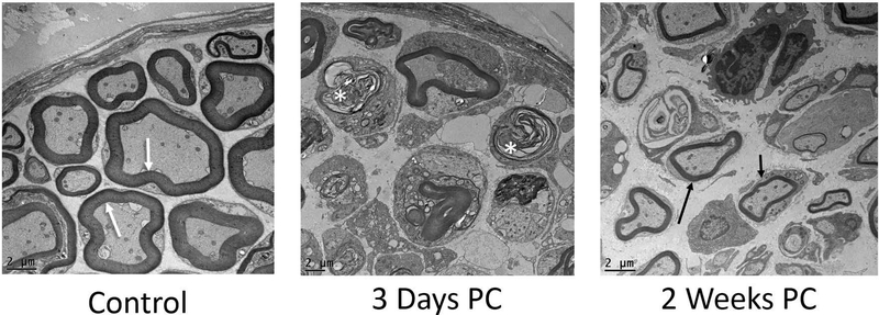
Figure 6:
Representative TEM images of control and experimental RLN (above; 1200x). Left (control) nerves showed thick myelination (white arrows) and tightly packed axons. At 3 days post-crush, the right (experimental) RLN showed extensive signs of degeneration, indicated by collapsed fibers and dense, compressed myelin debris (asterisks). At 2 weeks post-crush, regeneration of thinly myelinated axons was evident (arrows) within an expanded perineurial space.

