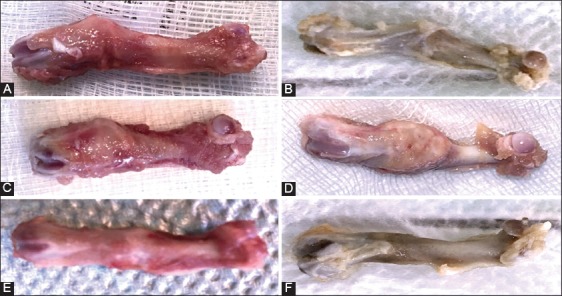
FIGURE 2.

Macroscopic findings in control and HF-PEMF group at two and eight weeks after surgery. A) There was a delay in consolidation in control group, but B) valgus consolidation was observed in HF-PEMF group. C) At two weeks, bone callus in control group was composed of fibrous tissue, while D) in HF-PEMF group it had calcified islands. E) At eight weeks in control group, the femur diameter at the fracture site was larger than the diameter of the adjacent proximal and distal regions. F) In HF-PEMF group, the diameter of the femur was almost normal at eight weeks. HF-PEMF: High-frequency pulsed electromagnetic field.
