
FIGURE 6.
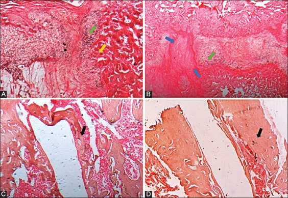
Histological analysis of femurs in control and HF-PEMF group at two and eight weeks after surgery (4×, hematoxylin and eosin [H&E]). A) At two weeks postoperatively, femur samples from control group showed a persistent infiltration of inflammatory cells (yellow arrow) and numerous chondrocytes (green arrow) at the fracture site. B) At eight weeks, there was a lower amount of bone marrow in the medullary cavity and less defined woven bone trabeculae (black arrow) in femurs from control group. C) In HF-PEMF group at two weeks, soft callus was at a more advanced, fibrocartilaginous stage and there was synthesis of new collagen fibers (blue arrow). These samples had no inflammatory infiltrates and had less chondrocytes (green arrow). D) At eight weeks, femurs from HF-PEMF group had a completely formed woven bone (black arrow) with dense trabeculae and had active bone marrow. HF-PEMF: High-frequency pulsed electromagnetic field.