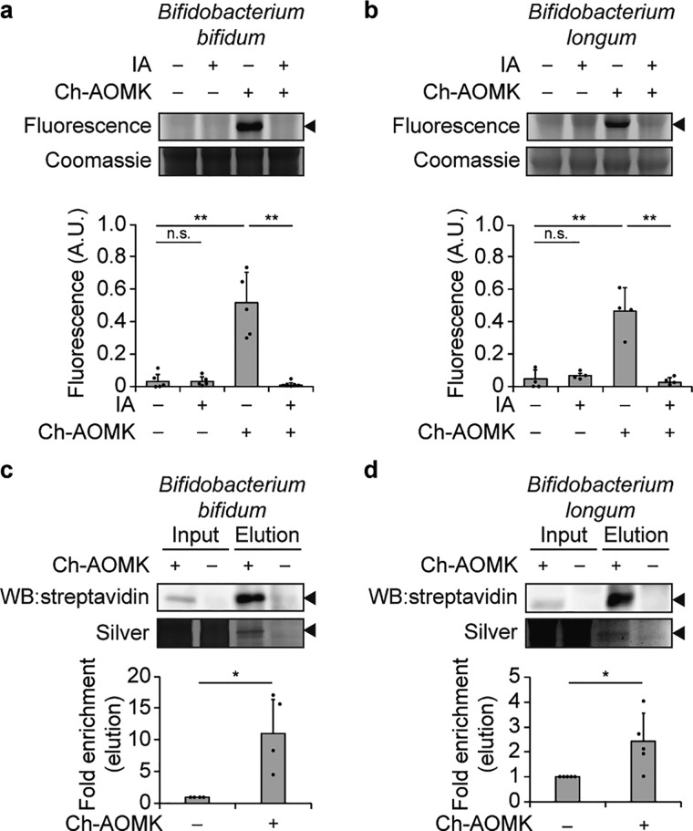
Figure 4.
Ch-AOMK labels BSH from gut anaerobes. Ch-AOMK (500 μM) was incubated with lysates from (a, c) Bifidobacterium bifidum or (b, d) Bifidobacterium longum at 37 °C for 24 h. (a, b) Lysates were treated with iodoacetamide (IA, 20 mM) prior to incubation with Ch-AOMK as a negative control. Following Ch-AOMK labeling, CuAAC tagging was carried out with (a, b) Fluor 488-alkyne or (c, d) biotin-alkyne. (a, b) Samples were subjected to SDS-PAGE, and the gel was visualized using fluorescence, followed by Coomassie staining. (c, d) Samples were analyzed either by Western blot with streptavidin-HRP or by silver staining. Input is 2% of the elution. The arrowhead indicates the expected size of BSHs (35 kDa). A.U. = arbitrary unit. The bands were quantified by densitometry using ImageJ (bottom panels). Error bars represent standard deviation from the mean. * p < 0.05, ** p < 0.01, *** p < 0.001, n.s. = not significant, n = (a) 5, (b) 4, (c) 4, (d) 5.

