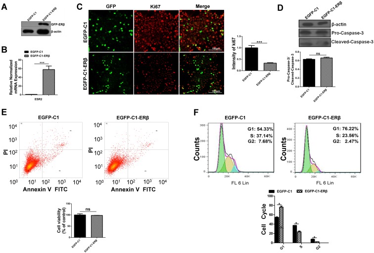
Figure 1.
ERβ plays an anti-proliferation role through impairing cell cycle but not apoptosis in HCT116 cells. HCT116 cells were transiently transfected with EGFP-C1 and EGFP-C1-ERβ plasmids for 48 h, and overexpression efficiency was determined by western blotting (A) and qRT-PCR (B). (C) Immunofluorescence analysis of HCT116 cells with Ki67 (red) and GFP (green), which were visualized by the confocal microscope. Bar graph (right) indicates the percentage of ki67 positive cells. Scale bar, 100μm. (D) Immunoblot analysis of pro-caspase-3 and cleaved-caspase-3. β-actin was used as an equal loading control. Bar graph (below) shows the relative ratio of pro-caspase-3 to cleaved-caspase-3 in HCT116. (E) Apoptosis was determined by flow cytometry after stained with Annexin V-FITC and PI. Bar graph (below) indicates the survival rate of treated cells. (F) Cell cycle distribution was analyzed by flow cytometer after DAPI stained. Bar graph (below) indicates the cell cycle phase distribution in treated cells. Immunofluorescence intensity and western blot were quantified using ImageJ software. Data shown are mean ± S.D. of three independent experiments. (*, P<0.05; **, P <0.01; ***, P<0.001).

