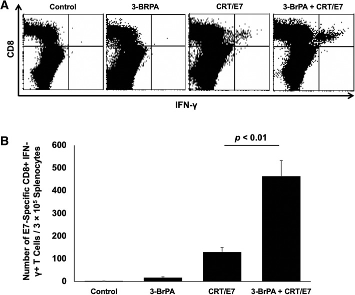
Fig. 2.
Characterization of E7-specific CD8+ T cells. Intracellular cytokine staining and flow cytometric analysis to determine the number of IFN-γ–secreting E7-specific CD8+ T cells in tumor-bearing mice treated with 3-BrPA and/or CRT/E7. On day 21, splenocytes from the treated TC-1 tumor-bearing mice were harvested and incubated with the E7 peptide overnight. Among complete splenocytes, E7-specific CD8+ T cells were quantified using intracellular staining for IFN-γ, followed by flow cytometry analysis. a Representative flow cytometric analyses data shown. b Bar graph depicting the number of E7-specific IFN-γ-producing CD8+ T cells per 3 × 10⁵ splenocytes. Each column represents the mean T cell count of each group; the standard deviation is indicated by the bars. Data is represented by the mean ± SD of three independent experiments

