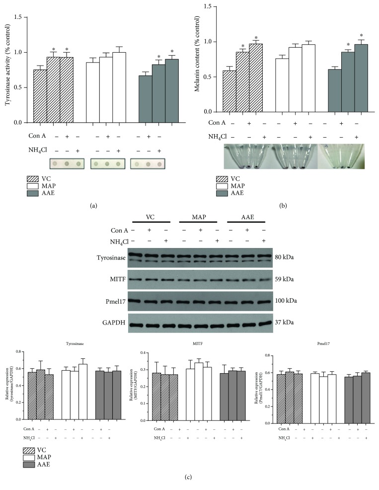
Figure 6.
Effect of intracellular neutralization on the tyrosinase activity, melanin content, and expression level of melanogenic proteins. MCs were untreated or treated with 1 mM VC, MAP, or AAE for 48 h; the tyrosinase activity (a) and melanin content (b) of treated MCs were measured by dot-blot assay and spectrophotometric analysis, as detailed in Materials and Methods. For pH neutralization, the MCs were simultaneously untreated or treated with 10 nM Con A or 10 mM NH4Cl. (c) The expression levels of tyrosinase, MITF, and Pmel17 proteins were analyzed by western blotting using anti-tyrosinase, anti-MITF, and anti-Pmel17 antibodies. GAPDH was used as a loading control. Representative blots are shown. Data are shown as means ± SD of three independent experiments. ∗ P < 0.05 versus only VC or its derivatives.

