Figure 8.
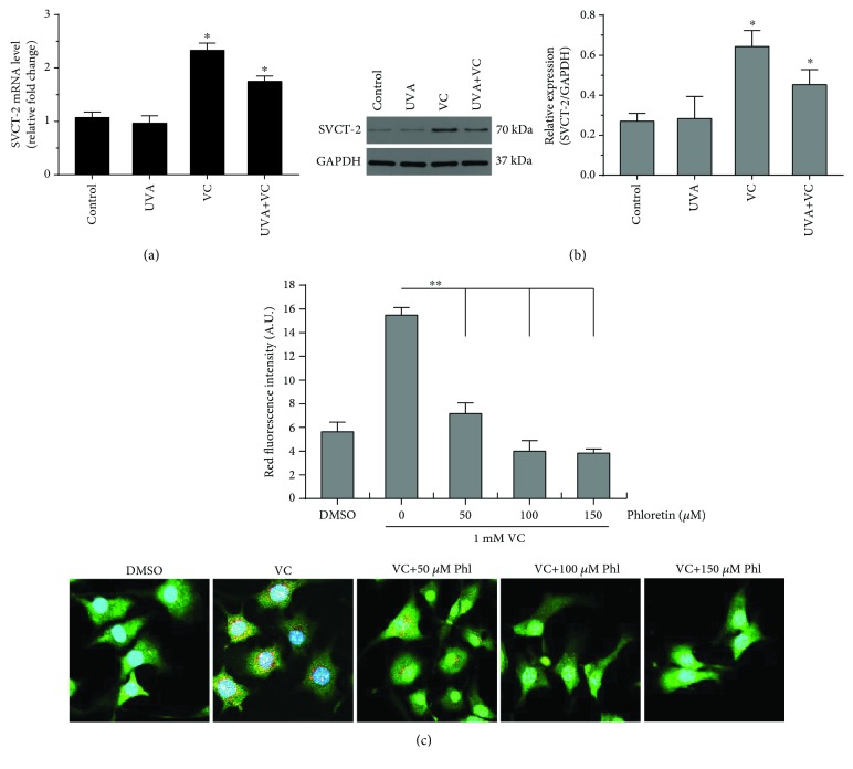
Expression levels of SVCT-2 mRNA and protein in MCs. MCs were seeded in 6-well plates and were then exposed to 3 J/cm2 UVA irradiation in the presence or absence of 1 mM VC. (a) The mRNA level of SVCT-2 was measured using qPCR as detailed in Materials and Methods. (b) The protein level of SVCT-2 was analyzed by western blotting using an anti-SVCT-2 antibody. GAPDH was used as a loading control. Representative blots are shown. Data are shown as means ± SD of three independent experiments. ∗ P < 0.05, compared to the untreated control. (c) For inhibiting SVCT-2, phloretin (a putative SVCT-2 inhibitor) was purchased from Selleck Chemicals (Cat# S2342, Shanghai, China). All compounds were first dissolved in dimethyl sulfoxide (DMSO) and then diluted with PBS into the indicated concentrations. The concentration of phloretin was confirmed to be nontoxic by a cell viability assay. Cells were treated and then stained with AO using the same procedure described in Figure 4. The equal volume of DMSO in PBS was used as a solvent control. Red fluorescence intensity (arbitrary units (A.U.)) for AO staining was measured using ImageJ. Histogram shows the results determined on 50 cells which are presented as means ± SD for three independent experiments. ∗∗ P < 0.01, versus only VC treatment. Representative immunofluorescence images are shown in the bottom. Scale bar: 10 μm.

