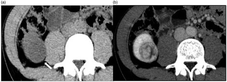
Fig. 2.
A 38-year-old man with clear cell RCC. (a) Unenhanced image; (b) contrast-enhanced image. On axial unenhanced CT, a 3.5-cm homogeneous low-density mass measuring 20 HU with a clear margin is detected in the right kidney (Rt. Kidney, arrow). The mass projects outside the renal cortex. It is easily detected, but apparently looks like a complicated cyst. On axial contrast-enhanced CT, the mass is heterogeneously enhanced. Therefore, this mass is suggested to be malignant tumor, such as RCC.

