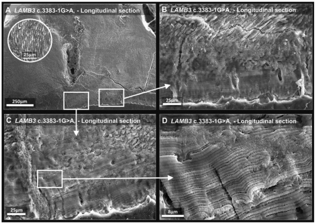
Figure 3.

Scanning electron microscopy of laminin subunit beta 3 (LAMB3) tooth 1. (A) Low-power image of affected enamel shows normal prismatic enamel in the bulk tissue away from the enamel-dentine junction; inset shows a higher-magnification image of the prismatic structure present. (B–D) The white squares indicate regions scanned at increasingly higher magnification. The initially secreted enamel, nearest the enamel-dentine junction, is aprismatic and appears to be composed of lamellae. For comparison, enamel from a control tooth is shown in Appendix Figure 4.
