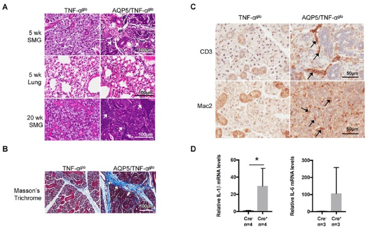
Figure 3.
Inflammation in the submandibular gland resulting from conditional overexpression of TNF-α. (A) Inflammatory infiltrates are seen in the submandibular glands of the AQP5/TNF-αglo mice at 5 wk of age (arrows), while other tissues, including the lungs, show no histologic signs of inflammation. A focus score of 90 ± 31 inflammatory infiltrates was counted per 200 × 200–μm square for male and female AQP5/TNF-αglo mice (total of 10 squares counted), while no inflammation was seen in the Cre- controls. By wk 20, severe inflammation is present in the submandibular glands, leading to glandular destruction (inflammatory foci were too numerous to count). (B) Chronic inflammation results in interstitial fibrosis, as seen by the abnormal collagen deposition shown by Masson trichrome staining. (C) CD3 staining shows recruitment of T cells in the submandibular glands of the AQP5/TNF-αglo mice. Mac2 staining shows an accompanying infiltration of macrophages into the submandibular glands around the blood vessels and ducts. (D) Additional proinflammatory cytokines are induced by overexpression of TNF-α in the submandibular gland, including a significant increase in IL-1β (*P ≤ 0.05, n = 4) and a trend toward increased IL-6 (n = 4). Values are presented as mean ± SEM. AQP5, aquaporin 5; SMG, submandibular gland.

