Figure 4.
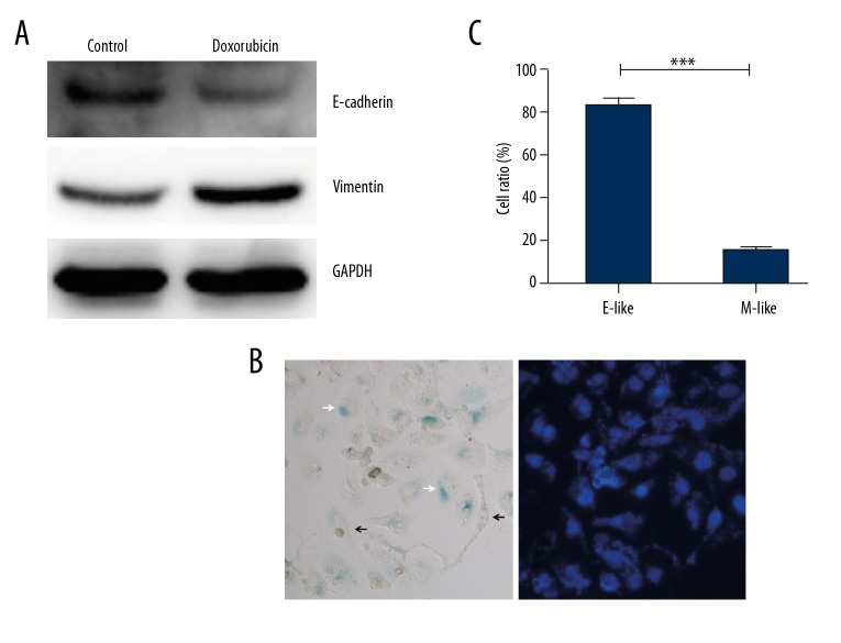
Morphological changes of senescence cells induced by doxorubicin. HeLa cells were treated with 0.1 μg/ml doxorubicin for 3 days followed by prolonged culture in drug-free medium for 6 days. (A) Expression of E-Cadherin and Vimentin were determined by Western blot. (B) After staining with SA-β-Gal kit and DAPI, most of the senescent cells showed an epithelial-like shape (white arrow), while mesenchymal-like cells were mainly SA-β-Gal-negative (black arrow). (C) Proportions of SA-β-Gal positive cells in epithelial(E)-like or mesenchymal(M)-like cells. *** p<0.001.

