Figure 1.

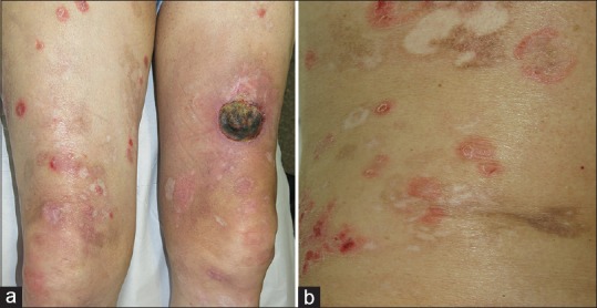
(a) Multiple papulonodular erythematous lesions and tumour with crustal surface located in lower extremities. (b) Some lesions had arcuate morphology and left residual hyper- and hypopigmentation

(a) Multiple papulonodular erythematous lesions and tumour with crustal surface located in lower extremities. (b) Some lesions had arcuate morphology and left residual hyper- and hypopigmentation