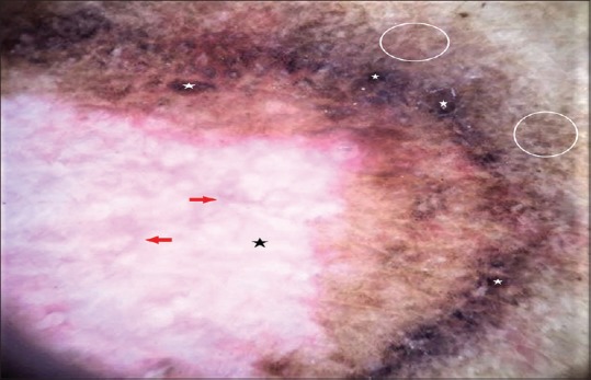
Figure 3.

Non-contact dermoscopy of the marginal area under polarized mode using DermLite™ DL3 shows dark globules (white stars) and irregular pigment network (white circles) surrounding a milky white area (black star) interspersed with fine telangiectatic vessels (red arrows) (original magnification ×10)
