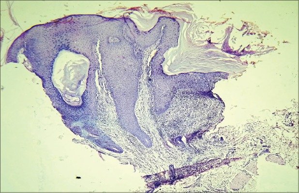
Figure 4.

Photomicrograph showing large invaginating masses of keratinocytes with overlying compact hyperkeratosis, hypergranulosis, and acanthosis. Also note the dilated follicular infundibulum with acanthosis and hypergranulosis of its epithelium (hematoxylin and eosin, original magnification ×5)
