Figure 2.

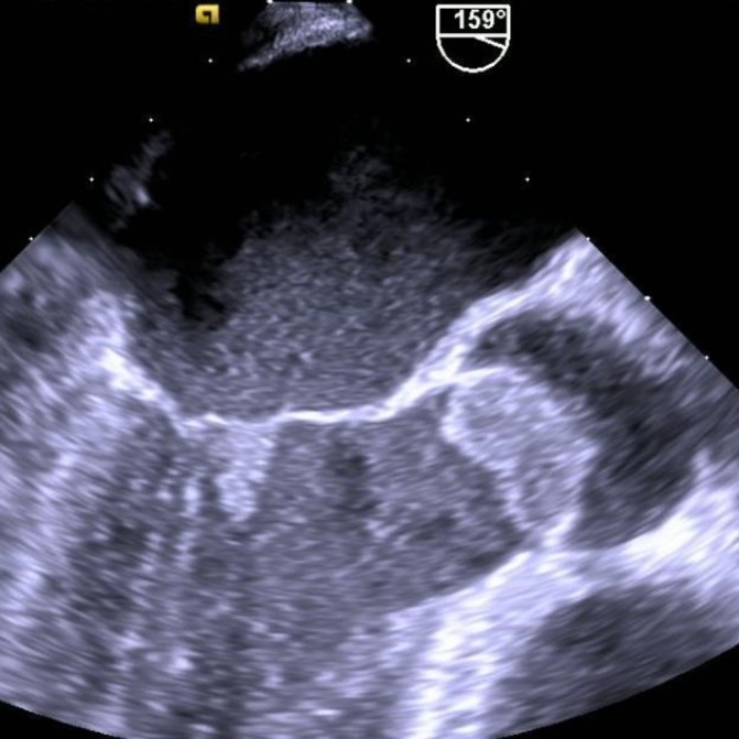
Transoesophageal echocardiographic images showing a large 19×16 mm vegetation attached to two and possibly all three aortic valve leaflets and a smaller 11×9 mm vegetation attached to the ventricular surface of the anterior mitral leaflet.

Transoesophageal echocardiographic images showing a large 19×16 mm vegetation attached to two and possibly all three aortic valve leaflets and a smaller 11×9 mm vegetation attached to the ventricular surface of the anterior mitral leaflet.