Figure 3.

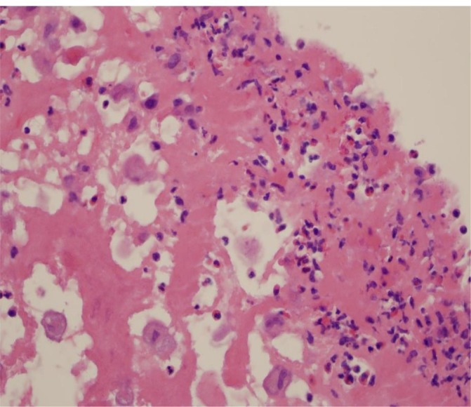
Histological section of the aortic valve vegetation showing areas of necrosis, histiocytes, multilobulated giant cells and eosinophils (H&E stain).

Histological section of the aortic valve vegetation showing areas of necrosis, histiocytes, multilobulated giant cells and eosinophils (H&E stain).