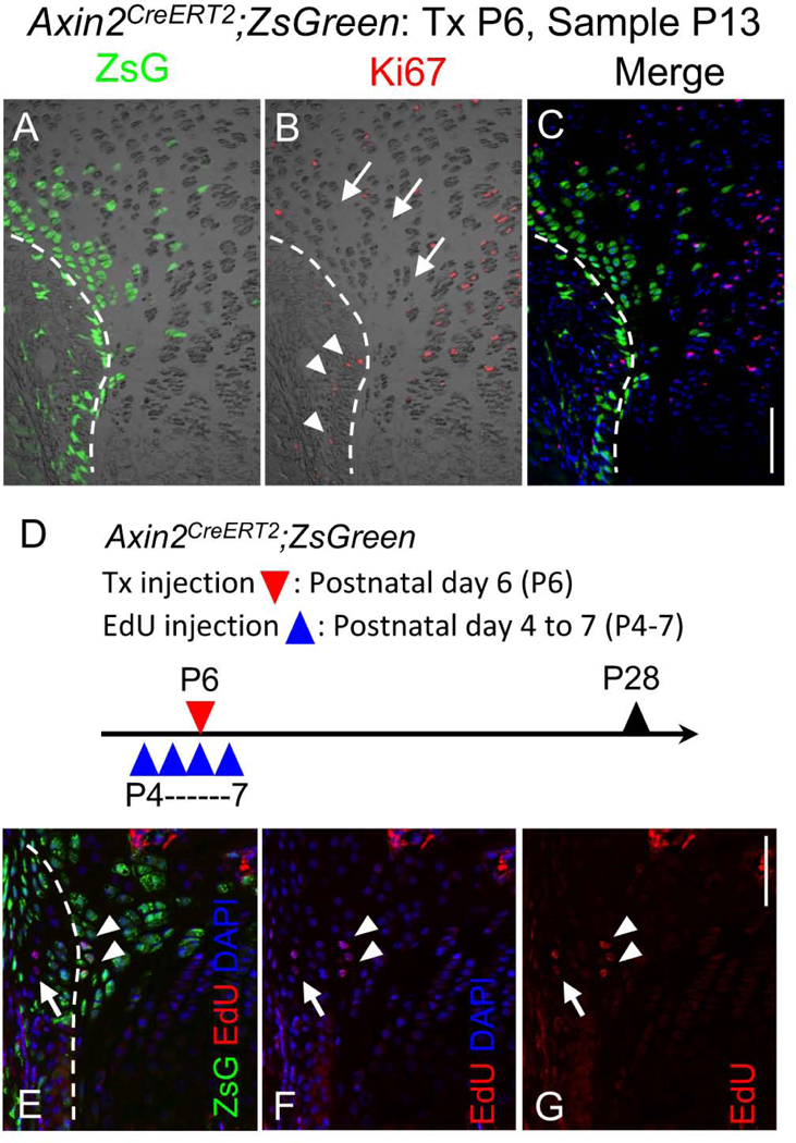
Fig. 3. Cell proliferation and slow-cycling cells of Wnt-responsive cells around the Ranvier’s groove.

(A-C) Immunohistochemical analysis for cell proliferation marker Ki67 protein on the proximal tibia GP sections of P13 female Axin2CreERT2;ZsG mouse that had received Tx injection at P6. A, ZsG (green) and DIC, B, Ki67 immunostaining (red) and DIC, C, Ki67 immunostaining (red), ZsG (green) and DAPI (blue). A small number of Ki67 positive cells were found in the Ranvier’s groove (arrow heads). The ZsG+ cells facing to the perichondrium showed no immunoreactivity for Ki67 antibody (arrows). Bar = 100 μm. (D-G) Long-term labeled cells with EdU in ZsG+ cells. D, Female Axin2CreERT2;ZsG mice received 4 daily intraperitoneal injections of EdU (5 μg/10 μl/mouse) from P4. Tx was injected at P6. Tibia were harvested at P28 and sections were subjected to EdU staining. E, ZsG (green), EdU (red) and DAPI (blue). F, EdU (red) and DAPI (blue). G, EdU (red). EdU-labeled cells were detected in Ranvier’s groove (arrows) and in the ZsG+ cells in the outermost layer of the GP (arrowheads). Bar = 100 μm.
