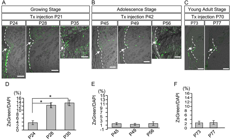
Fig. 5. Fate of Wnt-responsive cells around the Ranvier’s groove at different growth stages.

(A-C) Female mice carrying Axin2CreERT2;ZsG received Tx injection at P21 (growing stage, A), P42 (adolescence stage, B) or at P70 (young adult stage, C). The distribution of ZsG+ cells were histologically examined in the vicinity of RG after 3 days (P24, P45 or P73), 7 days (P28, P49 or P77), and 14 days (P35 or P56) post injection. Arrows represent the RG, and dotted lines indicate the border between perichondrium and growth plate. (D-F) Changes in the percentage of number of ZsG+ cells to number of DAPI positive cells in the growth plate within 200 μm range adjacent to the RG of each stage were examined. The values and error bars represent average and standard deviations. Note the significant increase of ZsG+ cells in the RG was only seen in growing stage (D). *, p < 0.05. Bar = 100 μm.
