
Fig. 6. Inactivation of β-catenin in Wnt-responsive cells leads to ectopic cartilage formation.
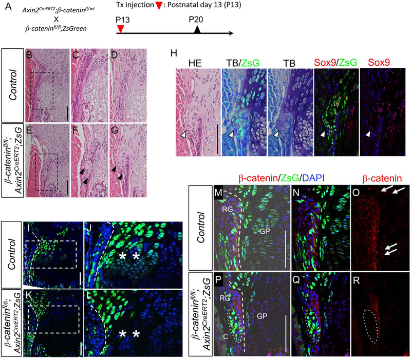
(A) β-cateninfl/fl mice possessing loxP sites in introns 1 and 6 in the β-catenin gene. Axin2CreERT2;β-cateninfl/wt were crossed with β-cateninfl/fl;ZsGreen. The resulting triple compound β-cateninfl/fl;Axin2CreERT2;ZsG mice received Tx injection at P13. Tibiae were harvested at P20. (B-G) Representative HE images of proximal tibias harvested from female mice. B-D, β-cateninfl/wt;Axin2CreERT2;ZsG (Control). E-G, β-cateninfl/fl;Axin2CreERT; ZsG. The images from two independent control (C and D) and target (F and G) mice were shown. In the β-cateninfl/fl;Axin2CreERT2;ZsG mice, the ectopic cartilaginous masses were found (arrowheads) in the groove of Ranvier (C and D). C and F are magnified images shown in squares of B and E, respectively. (H) Immunohistochemical and histochemical analysis of ectopic cartilaginous mass. HE, HE staining. TB/ZsG, Toluidin blue staining and ZsG (green). TB, Toluidin blue staining. Sox9/ZsG, Sox9 immunostaining (red), ZsG (green) abd DAPI (blue). Sox9, Sox9 immunostaining (red) and DAPI (blue). TB and TB/ZsG images were captured from the same section, and Sox9 and Sox9/ZsG images were captured from the serial section of the TB and TB/ZsG section. Note the ectopic cartilage shows cartilage matrix (stained purple) with TB and nuclear Sox9 protein expression. (I-L) Representative fluorescent images of the growth plate of proximal tibiae. I and J, β-cateninfl/wt;Axin2CreERT2;ZsG (Control). K and L, β-cateninfl/fl;Axin2CreERT2;ZsG. Note that the β-catenin-deficient mutants showed less expansion of ZsG+ cells toward the growth plate compared to the control mice (asterisks). Periphery of the growth plate (broken line) is shown in higher magnification in the right panels. Dotted line: the margin between RG and growth plate. (M-R) Efficiency of the β-catenin ablation was confirmed with immunohistochemistry using anti-β-catenin antibody. M-O, β-cateninfl/wt;Axin2CreERT2;ZsG (Control). P-R, β-cateninfl/fl;Axin2CreERT;ZsG. Note that the β-catenin-deficient mutants showed lower β-catenin protein expressions (arrowheads) compare to control mice. In the control mice, nuclear β-catenin expression was observed in the outermost cell layer of the growth plate (arrows). Bar = 100 μm.