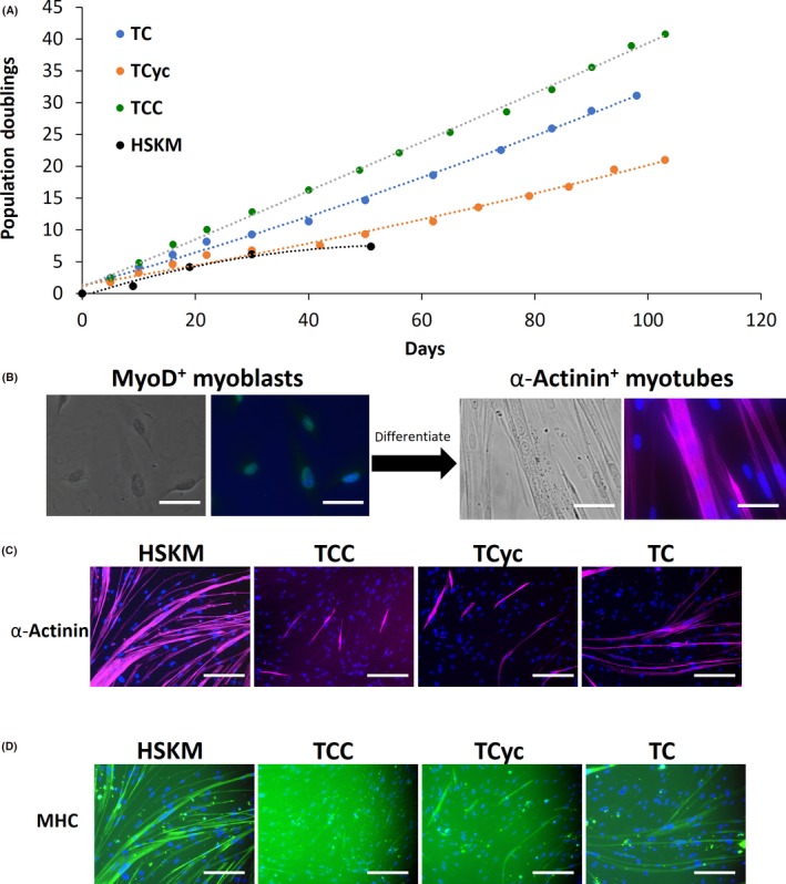
Figure 1.

Immortalization of adult primary human skeletal muscle (HSKM) myoblasts. A, Population doubling curves for HSKM myoblasts (black), and HSKM myoblasts transduced with lentiviral hTERT and Cyclin D1 (TCyc, orange), or CDK4R24C (TC, blue), or CDK4R24C and Cyclin D1 (TCC, green). While adult HSKM myoblasts underwent senescence by the 6th population doubling at 30 d, the other cells continued to proliferate steadily for 100 d and beyond. B, High magnification (40×) phase contrast and immunofluorescence images of MyoD+ (green) myoblasts and, after fusion and differentiation, α‐actinin+ (purple) multinucleated myotubes. Cells were counterstained with DAPI to visualize the myonuclei. Scale bars 25 μm. C, Immunofluorescence staining for the myotube marker α‐actinin in HSKM, TCC (hTERT, CDK4R24C, Cyclin D1), hTERT‐Cyclin D1 and hTERT‐CDK4 myoblasts that were subjected to myogenic differentiation. Cells were counterstained with DAPI to visualize the myonuclei. Scale bars 50 μm. D, Immunofluorescence staining for the myotube marker myosin heavy chain (MHC) in HSKM, TCC (hTERT, CDK4R24C, Cyclin D1), hTERT‐Cyclin D1 and hTERT‐CDK4 myoblasts that were subjected to myogenic differentiation. Cells were counterstained with DAPI to visualize the myonuclei. Scale bars 50 μm
