Figure 3.

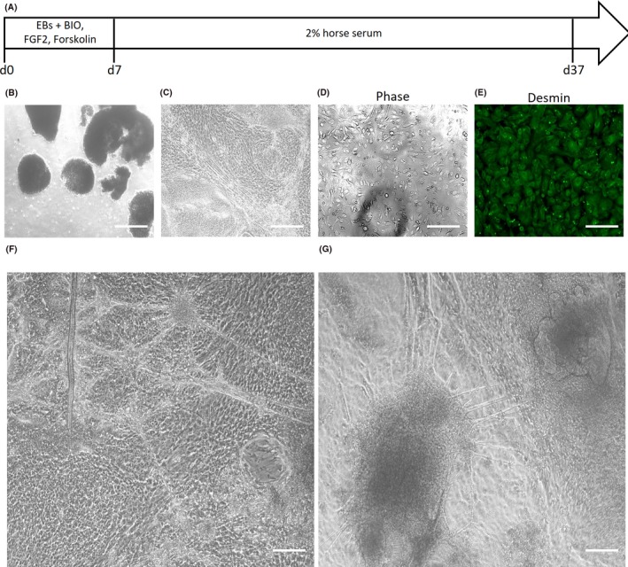
Derivation of human myoblasts and myotubes from highly proliferative human embryonic stem cells (hESCs) via embryoid bodies. A, Schematic of directed differentiation protocol for embryoid body (EB)‐derived myoblasts. B, Phase contrast micrographs of EBs derived from hESCs. Scale bar 200 μm. C, Phase contrast micrographs of mesodermal progenitors cultured from the hESC‐EBs. Scale bar 50 μm. D, Phase contrast micrographs, and (E) desmin immunofluorescence staining of heterogeneous myogenic cells derived from the hESC‐EB‐mesodermal progenitors. Scale bar 50 μm. F, Phase contrast micrograph of myotubes and myocytes derived from hESC‐EB‐myoblasts that were subjected to myogenic differentiation culture conditions for 2 wk. Scale bar 50 μm. G, Phase contrast micrograph of myotubes and myocytes derived from hESC‐EB‐myoblasts that were subjected to myogenic differentiation culture conditions for 3 mo. Scale bar 50 μm
