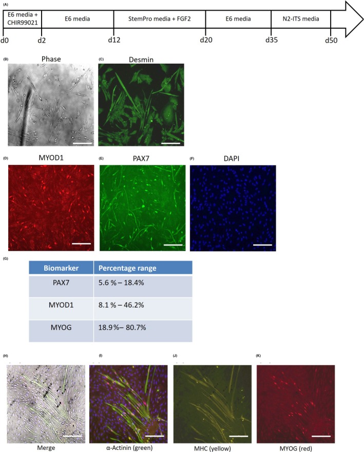
Figure 6.

Derivation of human myoblasts and myotubes from highly proliferative human embryonic stem cells (hESCs) via mesodermal monolayers. A, Schematic of directed differentiation protocol for mesodermal monolayer‐derived myoblasts. B, Phase contrast micrographs, and (C) desmin immunofluorescence staining of myogenic cells derived from the hESC‐mesodermal monolayer cultures. Scale bars 50 μm. D‐F, Immunofluorescence staining of the hESC‐mesodermal monolayer‐myogenic cells for the myoblast markers (D) MYOD1 (red) and (E) PAX7 (green), with nuclei counterstained by (F) DAPI (blue). Scale bars 100 μm. G, Quantification of PAX7+, MYOD1+ and MYOG+ cells amongst the hESC‐mesodermal monolayer‐myogenic cells. H, Immunofluorescence staining for (I) α‐actinin (green), (J) myosin heavy chain (MHC, yellow) and (K) myogenin (MYOG, red) in hESC‐monolayer‐myoblasts that were subjected to myogenic differentiation culture conditions for 2 wk. Scale bars 50 μm
