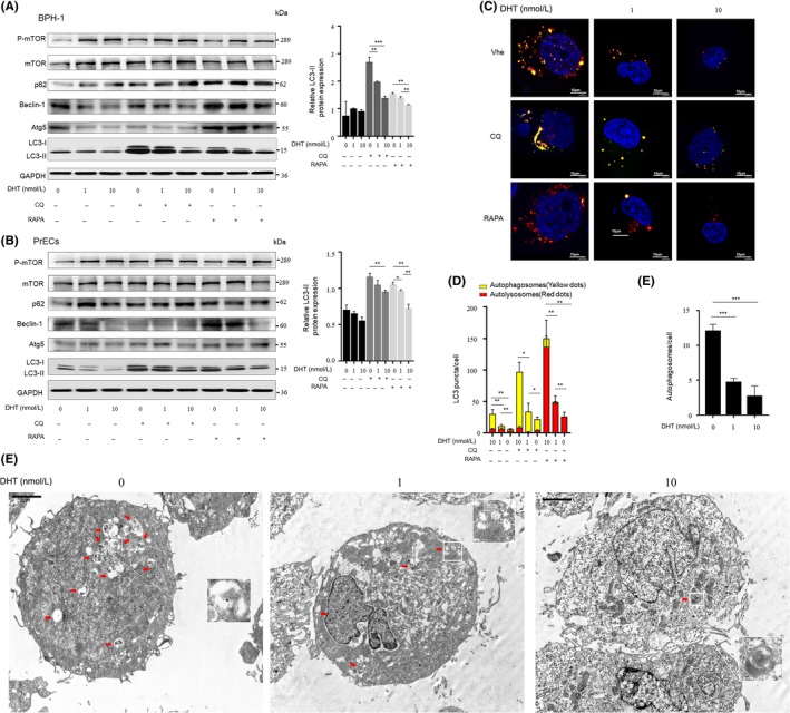
Figure 4.

Androgen ablation in prostate fibroblasts promotes autophagic flux in co‐cultured prostate epithelial cells. A and B, BPH‐1 or PrECs cells were treated with conditioned medium from WPMY‐1‐AR cells that were fed for 72 h with 0, 1 or 10 nmol/L DHT, respectively, followed by 50 nmol/L CQ or 50 μmol/L RAPA treatment for another 1 h. Left, Western blotting analysis. Right, statistical analysis of relative LC3‐II expression. C, BPH‐1 cells stably expressing an mRFP‐GFP‐LC3 fusion were treated with conditioned medium for 1 h and then fed with 50 nmol/L RAPA or 50 μmol/L CQ for another 1 h. (scale bars, 10 μm). Veh: vehicle control group. D, Average number of autophagosomes and autolysosomes in BPH‐1 was analysed and plotted. E, TEM results of BPH‐1 cultured with conditioned medium for 1 h before fixation (scale bars: 2 μm) (F) Quantification of the number of autophagic vacuoles. *P < 0.05, **P < 0.01, ***P < 0.001
