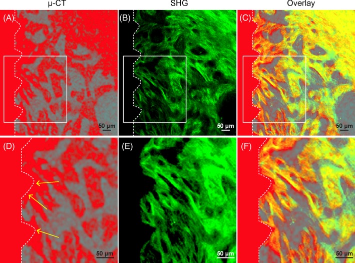
Figure 2.

Second harmonic generation (SHG) signal presents intact bone microstructure at the implant‐bone interface without artefact. Wild‐type mice were used for implant placement. Mandible samples were collected 1 mo after placement and processed following polyethylene glycol (PEG)‐associated solvent system clearing protocol with decalcification treatment. A, μCT image of the bone‐implant interface acquired before decalcification. High‐density bone and implant were shown in red colour. B, SHG signal (green) was acquired after clearing with a two‐photon microscope at identical position as in panel A. C, Overlay of (A) and (B). D‐F, Boxed area in (A), (B) and (C) were enlarged. Arrows in d indicate halation artefact of μCT. Scale bars, 50 μm
