
Figure 3.
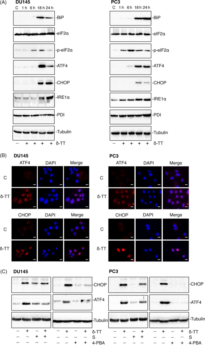
δ‐TT triggers ER stress in DU145 and PC3 prostate cancer cells. A, DU145 and PC3 cells were treated with δ‐TT (15 μg/mL) for 1‐24 h. Western blot analysis was performed to investigate the expression levels of ER stress‐related proteins (BiP, eIF2α, p‐ eIF2α, ATF4, CHOP, IRE1α, PDI). Tubulin expression was evaluated as a loading control. B, DU145 and PC3 cells were treated with δ‐TT (15 μg/mL) for 18 h. The expression levels and intracellular localization of the key transcription factors involved in the ER stress‐mediated apoptosis (ATF4 and CHOP) were evaluated by immunofluorescence analysis. C, controls (vehicle). Scale bars are 20 μm. C, CRPC cells were pretreated with the ER stress inhibitors salubrinal (S; 20 μM) or 4‐PBA (2 mM), for 4 and 1 h, respectively, before treatment with δ‐TT (15 μg/mL) for 24 h. The effects of the treatments were analysed on CHOP and ATF expression levels by Western blot. Tubulin expression was evaluated as a loading control. One representative of three different experiments performed is shown