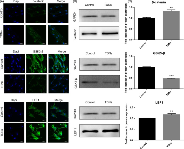
Figure 5.

The potential mechanism through which TDN regulated the osteogenic differentiation of PDLSCs. A, Immunofluorescent micrographs of β‐catenin protein, GSK3‐β protein and LEF1 protein. (nucleus: blue, β‐catenin, GSK3‐β protein and LEF1 protein: green). Scale bars are 25 µm. B, WB analysis of the β‐catenin, GSK3‐β and LEF 1 proteins expression. C, Statistical analysis of WB. Data are presented as means ± standard deviations (n = 3). ***P < 0.001.
