Figure 5.

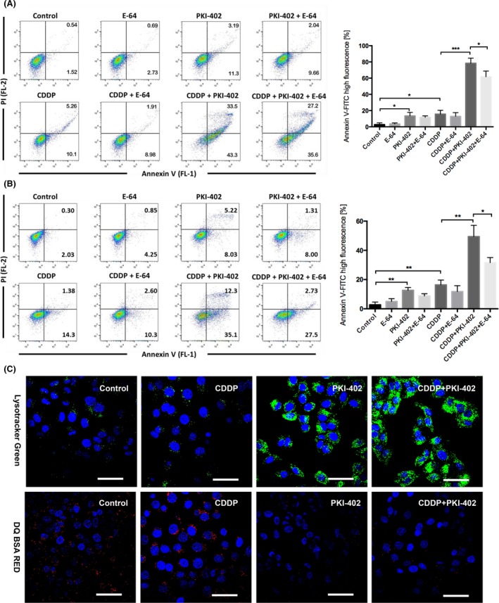
PKI‐402 combined with cisplatin significantly increased apoptosis and induced lysosomal dysfunction in HCC cells. A, Huh7 cells were treated with 8 μg/mL cisplatin and/or 5 μmol/L PKI‐402 in the presence or absence of 5 μmol/L E‐64 for 24 h and then stained with Annexin V and PI and analysed by FlowJo. The percentage of cells with high Annexin V‐FITC fluorescence is expressed as the mean ± SD; n = 3, *P < 0.05, ***P < 0.001. B, HepG2 cells were treated with 12 μg/mL cisplatin and/or 5 μmol/L PKI‐402 in the presence or absence of 5 μmol/L E‐64 for 24 h and then stained with Annexin V and PI and analysed by FlowJo. The percentage of cells with high Annexin V‐FITC fluorescence is expressed as the mean ± SD; n = 3, *P < 0.05, **P < 0.01. C, Huh7 cells were treated with 8 μg/mL cisplatin and/or 5 μmol/L PKI‐402 for 12 h and then stained with LysoTracker Green DND‐26 and DQ Red BSA and observed with confocal laser microscopy (scale bar = 20 μm)
