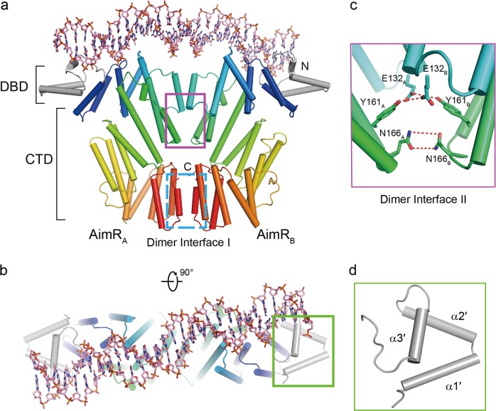
Fig. 1. Structure of AimR–DNA complex.
a, b The overall structure of AimR–DNA. The CTDs of AimR are rainbow-colored and the DBDs of AimR are colored in gray. The DNA fragment (5′ CTTAAATATTAGGTTTTAATAACATCTAGT 3′) is shown by stick. Two dimer interfaces between the two protomers of AimR are highlighted using rectangle. c Close-up view of dimer interface II. The hydrogen bonds are represented by red dotted lines. d The α-helices are sequentially numbered from the N terminus of the DBD

