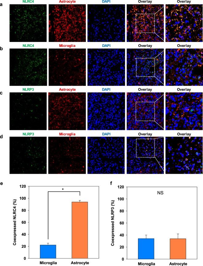
Figure 3.
NLRC4 is highly expressed in astrocytes from glioma patients. Astrocytes (a,c) and microglia (b,d) were analyzed by immunohistochemistry for the expression of NLRC4 and NLRP3, respectively. Nuclei were stained with DAPI (blue). Co-expression of NLRC4 (e) or NLRP3 (f) with markers for microglia and astrocytes was quantified by ImageJ software. The values are means ± SEMs from three replicates (n = 8). *T-test p-value < 0.001.

