
Figure 6.
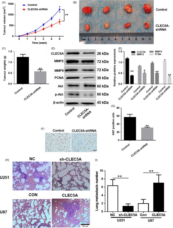
Knockdown of CLEC5A inhibited tumour growth in vivo. A, Time‐dependent tumour volume of cell transplantation in nude mice with shCLEC5A U251 normalized to control cells. B, After 35 d incubation of shCLEC5A transfected U251 cells in mice, the tumour was isolated and pictured. C, Quantification of tumour weight after 35 d of cell transplantation in nude mice with shCLEC5A U251 normalized to control cells. D, Protein levels of PCNA, MMP2, MMP9, Akt and Akt phosphorylation in tumour tissues were detected by Western blot. E, Quantification of Western blot of protein levels of PCNA, MMP2, MMP9, Akt and Akt phosphorylation. F and G, Immunochemistry staining and quantification of Ki67 in tumour sections. H and I, Lung metastasis staining and quantification on mice tail vein injection of CLEC5A knocking down U251 or overexpressed U87 cells, respectively. Values are expressed as means ± SD, *P < 0.05, **P < 0.01 as compared to the control