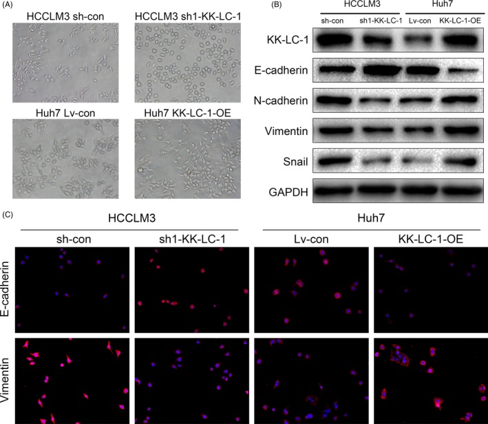
Figure 4.

KK‐LC‐1 is associated with EMT process in HCC. A, Representative images of cellular morphology of sh1‐KK‐LC, KK‐LC‐OE and their control cells. B, Expression levels of EMT markers mediated by KK‐LC‐1 were detected by Western blot. C, Immunofluorescence for E‐cadherin and vimentin showed that KK‐LC‐1 affected expressions of EMT markers. Representative images of sh1‐KK‐LC, KK‐LC‐OE and their control cells are shown
