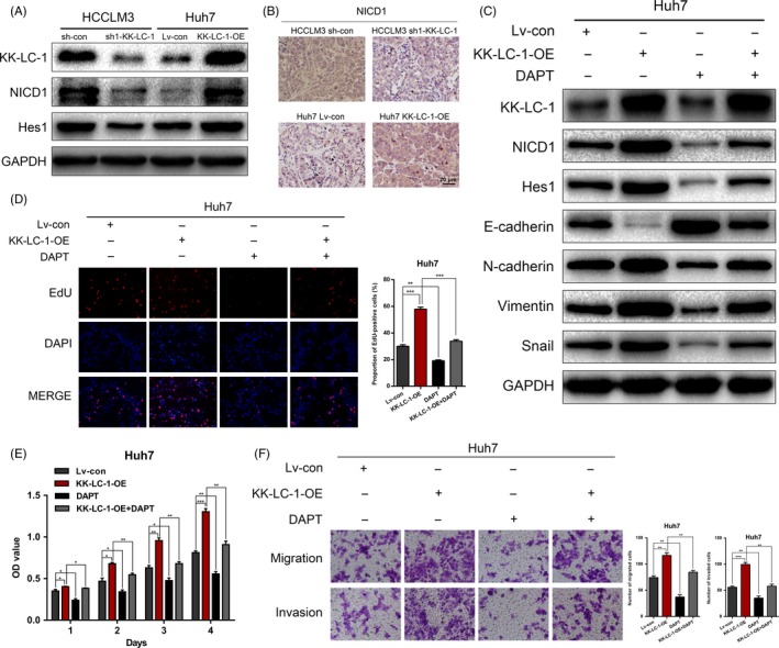
Figure 7.

KK‐LC‐1 activates the Notch1 signalling in HCC. A, Protein expressions of KK‐LC‐1, NICD1 and Hes1 were detected in HCCLM3 sh1‐KK‐LC‐1, Huh7 KK‐LC‐1‐OE and their control cells. B, Representative images of NICD1 immunostaining of the tumours derived from xenograft mice model. C, Western blot was performed to evaluate the expressions of NICD1, Hes1 and EMT markers in Huh7 KK‐LC‐1‐OE cells treated with DAPT. D, EdU assays were conducted to assess the proliferation ability of Huh7 KK‐LC‐1‐OE cells treated with DAPT. Proportion of EdU‐positive cells was determined and statistically analysed. E, CCK‐8 assays for Huh7 KK‐LC‐1‐OE cells treated with DAPT. F, Transwell assays were used to analyse HCC cell migration and invasion in Huh7 KK‐LC‐1‐OE cells treated with DAPT. Migrated and invaded cells were quantified and statistically analysed. The data are shown as the mean ± SE. *P < 0.05, **P < 0.01.
