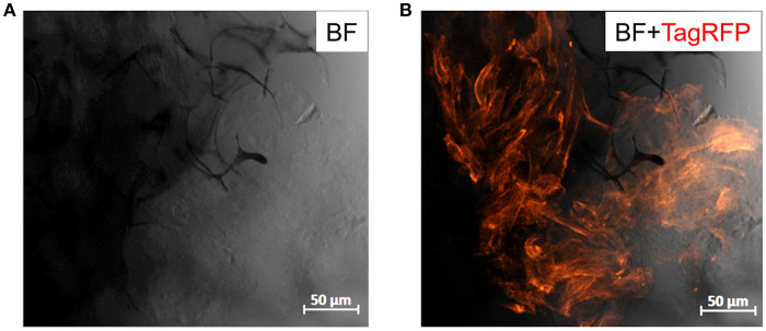
Figure 6.
Confocal images of PEDOT:PSS/MWCNTs (2:3) after 2 days in culture with TIF LifeAct cells. (A) Bright-field channel illustrating the scaffold porous network (dark gray-black) covered with cells (light gray), (B) Bright-field and far-red channels merged highlighting the actin filaments of TIF cytoskeleton (dark orange), as well as the cell growth and penetration into the pores of the scaffold.

