Figure 2.
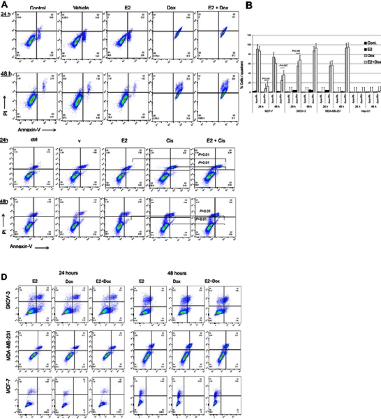
Levels of apoptosis in various cell types following treatment with E2 + doxorubicin (Dox) or cisplatin (Cis).
Notes: (A) Percentage apoptosis in MCF7 cells treated with 20 nM E2, 1 μg Dox, or a combination of both for 24 or 48 hours evaluated using annexin V–PI flow cytometry. (B) Means ± SD of percentage apoptosis in MCF7, SKOV3, MDA-MB231, and HepG2 cells treated with 20 nM E2, 1 μg Dox, or both for 24 or 48 h. Data based on three separate experiments. Percentage apoptosis in (C) A549 and (D) MCF7, SKOV3, and MDA-MB231 cells treated with E2 (20 nM), Cis (1 μg), or both for 24 or 48 hours.

