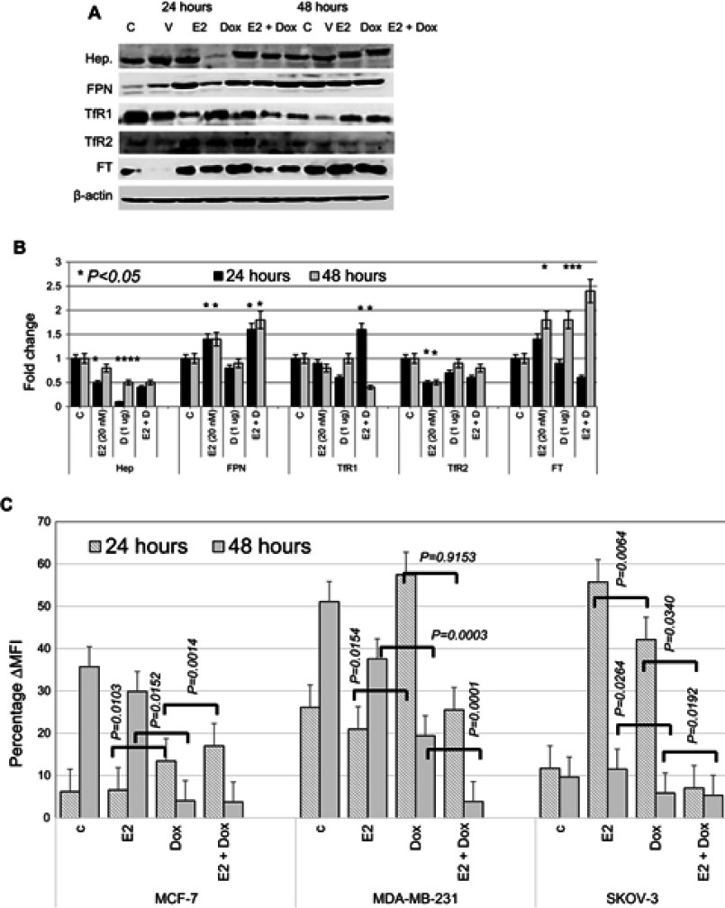
Figure 7.
Status of key proteins involved in iron metabolism and labile iron content in treated and control MCF7 cells.
Notes: (A) Representative Western blot of cell lysates prepared from MCF7 cells treated with E2, doxorubicin (Dox) or E2+Dox for 24 or 48 hours; the same nitrocellulose membrane was successively reacted with and stripped of secondary antibodies corresponding to the various proteins listed in the figure. (B) Calculated fold change ± SD of protein-expression levels in treated and control cells based on three separate experiments. (C) Labile iron content in MCF7 cells treated as in A using the CA-AM/CA-AM+DFO staining–based flow cytometry and expressed as %∆MFI.
Abbreviations: CA-AM, calcein acetoxymethyl; MFI, mean fluorescence intensity.

