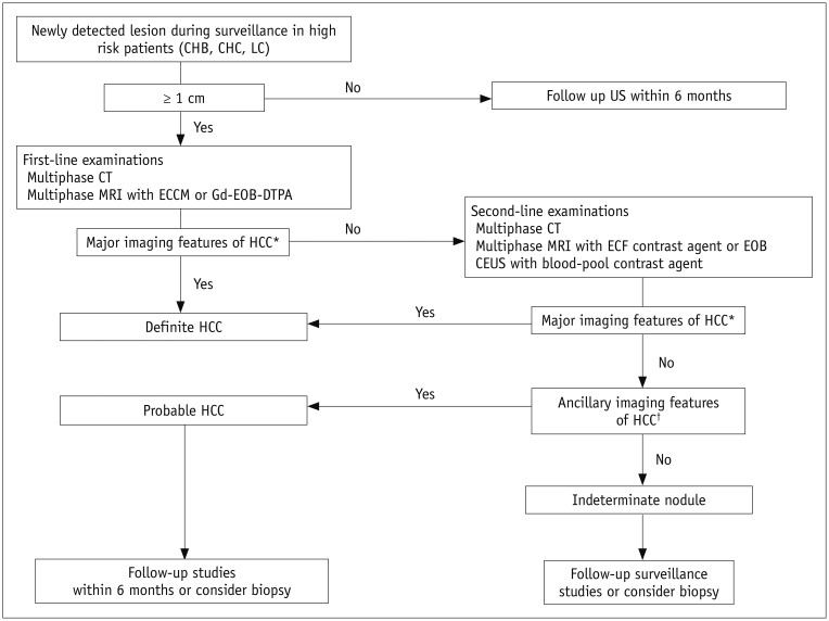
Fig. 3. Diagnostic algorithm for suspected HCC using new KLCA-NCC practice guidelines.
*Major imaging features of HCC include arterial hyperenhancement and washout appearance during portal venous, delayed, or HBP on multiphasic CT or MRI using extracellular contrast agents or EOB in nodules ≥ 1 cm in diameter. However, lesion should not show either marked T2 high SI or targetoid appearance on DWI or contrast-enhanced sequences. On CEUS as second-line examinations, major imaging features include arterial hyperenhancement and late (≥ 60 seconds) and mild washout, †In nodule(s) with some but not all aforementioned major imaging features of HCC, category of “probable” HCC can be assigned only when lesion fulfills at least one item from each of following two categories of ancillary imaging features. Two categories which make up ancillary imaging features are findings favoring malignancy in general (mild-to-moderate T2 hyperintensity, restricted diffusion, HBP hypointensity, interval growth) and those favoring HCC in particular (non-enhancing capsule, mosaic architecture, nodule-in-nodule appearance, fat or blood products in mass). These criteria should be applied only to lesion which shows neither marked T2 hyperintensity nor targetoid appearance on DWI or contrast-enhanced sequences. DWI = diffusion-weighted imaging, ECCM = extracellular contrast media, Gd-EOB-DTPA = gadolinium ethoxybenzyl diethylenetriamine pentaacetic acid, HBP = hepatobiliary phase, KLCA-NCC = Korean Liver Cancer Association-National Cancer Center, LC = liver cirrhosis, MRI = magnetic resonance imaging, SI = signal intensity

