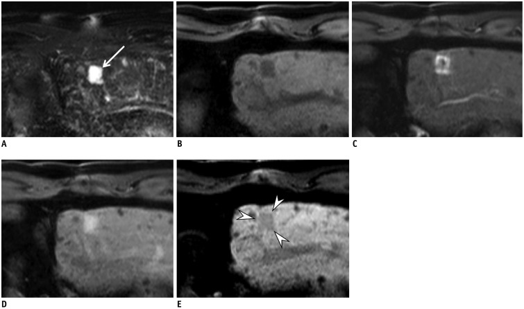
Fig. 5. Gadoxetic acid-enhanced MRI in 70-year-old man with CHB.
On T2-weighted (A) and precontrast T1-weighted (B) MRI, 2-cm nodule (arrow) is seen with marked T2 hyperintensity and low T1 SI at subcapsular portion of segment 2 of liver. After contrast injection, lesion demonstrates nodular enhancement in arterial phase (C) and persistent enhancement in portal phase (D). Lesion (arrowheads) depicts hypointensity on HBP (E). Lesion was proved to be hemangioma by showing no interval change over years. Regardless of arterial enhancement with hepatobiliary defect, diagnosis of HCC cannot be made due to exclusion criteria of marked T2 hyperintensity according to updated KLCA-NCC guidelines version 2018.

