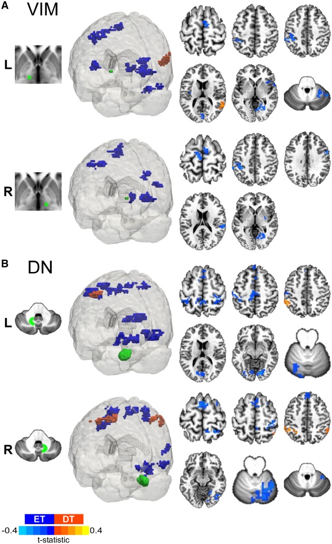
Figure 7.
Dystonic tremor versus essential tremor VIM and dentate nucleus FCΔ. 3D rendering and axial slice representation of whole-brain t-statistical maps comparing FCΔ of (A) VIM thalamus and (B) dentate nucleus (DN) for the dystonic tremor (DT) versus essential tremor (ET) comparison. Seed regions of interest are shown in green and the top and bottom panels depict the between-group FCΔ effects originating from the left (L) and right (R) seeds, respectively. In each panel, positive (red-orange) clusters denote significantly increased FCΔ with the seed region of interest in the dystonic tremor group compared to essential tremor group, whereas negative (blue) clusters denote significantly reduced FCΔ with the seed region of interest in the dystonic tremor group compared to essential tremor group (P < 0.05 FWER-corrected).

