Figure 2.
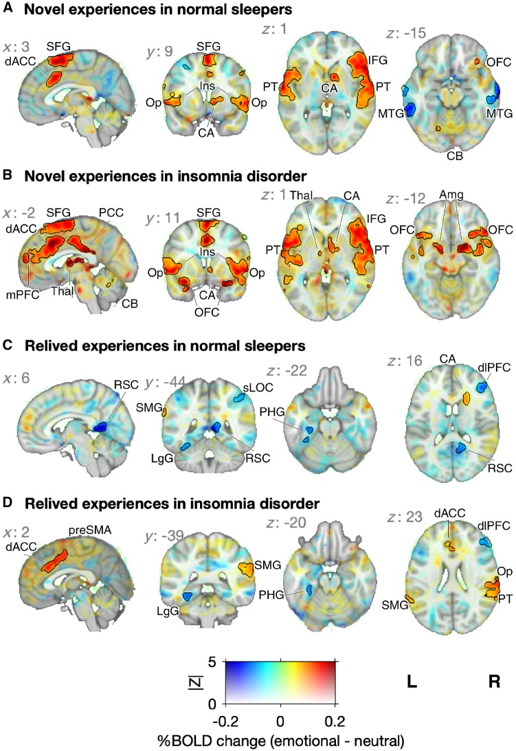
Emotion-specific BOLD responses to novel and relived experiences. Group means for the contrast between emotional and neutral stimuli. The mean percent BOLD change is coded by hue and the Z-statistic is coded by opacity. Significant clusters are indicated with black outlines (FLAME 1+2, |Z| > 3.10 and P < 0.05; see Supplementary Tables 5–8 for detailed description of clusters). (A) In normal sleepers, emotion-specific BOLD activations to novel experiences were observed in the salience network, the extended auditory system, and the orbitofrontal cortex. (B) In patients with insomnia, emotion-specific BOLD activations to novel experiences included these areas but covered wider areas and were also seen in the amygdala. (C) In normal sleepers, emotion-specific BOLD activations to relived experiences were observed in the left supramarginal gyrus and just anterior and superior of the caudate nucleus (D) whereas in patients with insomnia, BOLD activations were most pronounced in the dorsal ACC, and pre-SMA, right parietal operculum cortex, right planum temporale, right parietal operculum cortex, and bilateral supramarginal gyrus. Amg = amygdala; CA = caudate nucleus; CB = cerebellum; dACC = dorsal ACC; dlPFC = dorsolateral prefrontal cortex; IFG = inferior frontal gyrus; Ins = insula; LgG = lingual gyrus; mPFC = medial prefrontal cortex; MTG = middle temporal gyrus; OFC = orbital frontal gyrus; Op = operculum cortex; PCC = posterior cingulate cortex; PHG = parahippocampal gyrus; PT = planum temporale; RSC = retrosplenial cortex; SFG = superior frontal gyrus; sLOC = superior lateral occipital cortex; SMG = supramarginal gyrus; Thal = thalamus.

